Decoding Heart Rate Signals To Refine Brain Stimulation Therapies for Depression


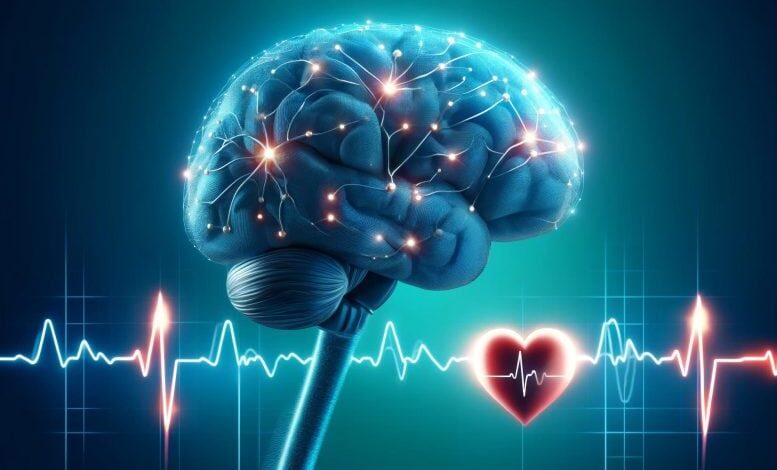
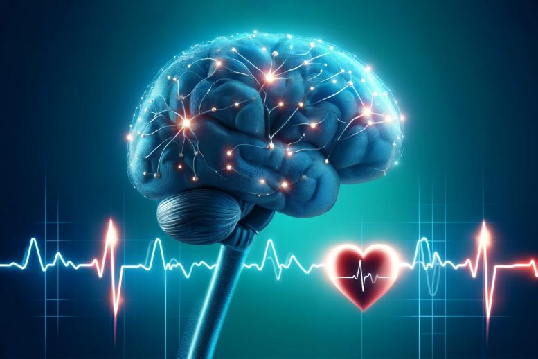
A study by Brigham and Women’s Hospital suggests that monitoring heart rate during transcranial magnetic stimulation (TMS) could help pinpoint effective stimulation sites in the brain for depression treatment, potentially eliminating the need for MRI scans. Credit: SciTechDaily.com
Study suggests heart rate may be a useful tool to determine where to stimulate the brains of individuals with depressive disorders when brain scans aren’t available.
New research suggests a common brain network exists between heart rate deceleration and depression. By evaluating data from 14 people with no depression symptoms, the team of researchers at Brigham and Women’s Hospital, a founding member of the Mass General Brigham healthcare system, found stimulating some parts of the brain linked to depression with transcranial magnetic stimulation (TMS), also affected heart rate, suggesting clinicians may be able to target those areas without the use of brain scans that aren’t widely available. The findings were published recently in the journal Nature Mental Health.
Heart-Brain Coupling and TMS
“Our goal was to figure out how to harness TMS treatment more effectively, get the dosing right, by selectively slowing down heart rate and identifying the individual best spot to stimulate on the brain,” said senior author Shan Siddiqi, MD, of the Brigham’s Department of Psychiatry and the Center for Brain Circuit Therapeutics. Siddiqi said the idea first developed during a conference in Croatia where researchers from the Netherlands were presenting heart-brain coupling data. “They showed that not only can TMS transiently lower the heart rate, but it matters where you stimulate,” Siddiqi said, adding that most exciting part of the study for him is the potential to give the rest of the world easier access to this precision targeted treatment for depression. “We have so many things we can do with advanced technology available here in Boston to help people with their symptoms,” he said. “But some of those things couldn’t easily get to the rest of the world before.”
Siddiqi collaborated with his colleagues in the Center for Brain Circuit Therapeutics at the Brigham and lead author, Eva Dijkstra, MSc, to complete the study. Dijkstra is a PhD candidate and came to the Brigham from the Netherlands to combine their work on heart-brain coupling with the CBCT team’s work on brain circuitry.
Research Methodology and Findings
Researchers looked at functional MRI scans from 14 people and identified spots in their brains believed to be optimal targets for depression based on previous studies done on connectivity and depression. Each participant had 10 spots identified in their brains, both optimal (‘connected areas’) and non-optimal for depression treatment. Researchers then observed what happened to heart rate when they stimulated each spot.
“We wanted to see if there would be mostly heart-brain coupling in the connected areas,” Dijkstra said. “For 12 out of 14 usable data sets, we found we would have a very high accuracy of defining an area that is connected by just measuring heart rate during brain stimulation.”
Dijkstra said the finding could help in both individualizing TMS therapy for depression treatment, by choosing a personalized treatment spot on the brain, and making it more accessible because an MRI wouldn’t need to be done beforehand.
Siddiqi said the findings from this study could also be used to help develop treatments that may eventually be useful to cardiologists and emergency doctors in a clinical setting.
Study Limitations and Future Research
One limitation of the study is it was done on a small number of people, and researchers didn’t stimulate all the possible spots in the brain.
The team’s next goal will be to map out where in the brain to stimulate to make heart rate changes more consistent.
Dijkstra’s team in the Netherlands is now working on a larger study of 150 people with depression disorders, many whose depression is treatment-resistant. Data from that study will be analyzed later this year, potentially bringing the research closer to clinical translation.
Reference: “Probing prefrontal-sgACC connectivity using TMS-induced heart–brain coupling” by Eva S. A. Dijkstra, Summer B. Frandsen, Hanneke van Dijk, Felix Duecker, Joseph J. Taylor, Alexander T. Sack, Martijn Arns and Shan H. Siddiqi, 26 April 2024, Nature Mental Health.
DOI: 10.1038/s44220-024-00248-8
Authorship: Additional authors include Summer B. Frandsen (BWH), Hanneke van Dijk, Felix Duecker, Joseph J. Taylor (BWH), Alexander T. Sack, and Martijn Arns.
Disclosures: Dijkstra is director and owner of Neurowave. Siddiqi has served as a speaker for Brainsway and PsychU.org (unbranded, sponsored by Otsuka). Siddiqi owns intellectual property involving the use of functional connectivity to target TMS. Additional disclosures can be found in the paper.
Funding: Brain and Behavior Research Foundation (Young Investigator Grant 31081), National Institutes of Health (K23MH129829, R01MH113929, R21MH12671, R21AA030372), the Baszucki Family Foundation, and the National Institute of Mental Health (PI: K23MH121657, Co-I: R01MH113929).

